Translate this page into:
Aviation Radiology: Teaching Series. Spinal Dysraphism
The basic events of central nervous system morphogenesis involve neurilation or the formation of the neural tube. The proximal two-thirds of the tube form the future brain and the caudal one - third form the spinal cord. The neural tube lumen becomes the ventricular system and the central canal of the spinal cord. The neural tube closes in a “zipper like fashion” beginning at the hindbrain and proceeding toward both ends of the embryo, Immediately after its closure, the superficial ectoderm of each side separates from the underlying neural ectoderm and then closes over it, this process being called dysjunction. The future meninges, neural arches, and paraspinal muscles are formed from mesenchyme that migrates dorsally between the neural tube and the skin. Premature separation or nondisjunction may result in severe anomalies. If the disjunction of the neural and cutaneous ectoderm occurs too early the adjacent mesenchyme can enter the neural tube resulting in anomalies like spinal cord lipoma. Focal failure of disjunction results in a persisting epithelial-lined communication between the ectoderm and the neural tube derivatives, i.e., a dermal sinus. A larger area of non-disjunction results in an open neural tube defect that is continuous dorsally with cutaneous ectoderm, i.e., myelocele or myelomeningocele.
The general term “spinal dysraphism” refers to those spinal anomalies that have incomplete midline closure of mesenchymal, osseous, and neural tissues.
Spinal Dysraphism can be broadly classified as :
(a) Open Spinal dysraphism
(b) Spina bifida
Open Spinal Dysraphism.
True breaches in the neural arch do, of course, occur, and they may be accompanied by a meningocele protruding posteriorly and usually in the lumbar region, this is known as Open Spinal Dysraphism. Occasionally, in the thorax and in the sacral region, the sac may protrude laterally and anteriorly through the intervertebral foramina and sacral foramina respectively. Other vertebral and rib anomalies are very frequent in the severe forms of spina bifida. The presence of hydrocephalus is a common feature in marked spina bifida.
Spina Bifida.
The neural tube normally closes at 3-4 weeks of gestational age. A failure of closure results in Spina Bifida. Lesions may occur anywhere along the spine but are commoner in the lumbar or lumbosacral regions (90%) than in the thoracic (6%) or cervical (3%) spines. The overall age-adjusted incidence is about 17%.
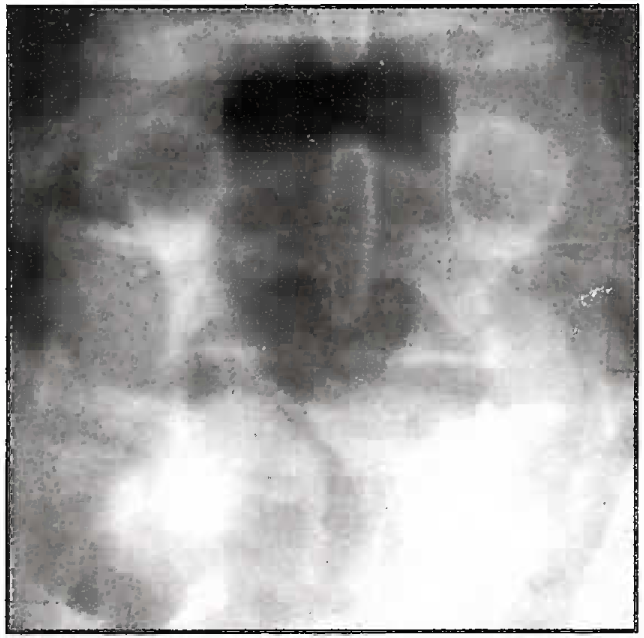
In most cases only a minor midsagittal defect in the neural arch is seen. The rather misleading term “Spina bifida occulta’ is applied to this condition. There is no true breach in such cases; the radiolucent area represents merely non-ossified cartilage. In children many of these areas become ossified as growth progresses.

- Spina Bifida
Spina bifida occulta is common. Even though there is a very slightly increased chance of early degenerative disc disease, very few people with spina bifida occulta will ever have any problems because of it. If a person has no symptoms from spina bifida occulta as a child, then it is unlikely that they will have any as an adult. Most people will not even be aware that they have spina bifida occulta unless it shows upon an X-ray which they have for some unrelated reason.
However, for some people (about 2% of those who have spina bifida occulta) there can be other problems. To avoid confusion, the term often used for spina bifida occulta with these associated problems is Occult Spinal Dysraphism.
Features of Occult Spinal Dysraphism (OSD) are:
Detrusor overactivity in children
Lipomas in the spine, under the skin or in surrounding tissues
Cysts in the skin or just under it
Syrinxes in the spine
Divisions in the spinal cord
Tethering of the spinal cord.
The normal spinal cord moves freely in the spinal canal. However, sometimes in OSD, the cord becomes tethered or stuck down. This can cause stretching of the cord and affect the blood flow to the area, especially during times of rapid growth.
Some of the symptoms of a tethered spinal cord are:
Motor weakness
Increased muscle tone
Deterioration in gait
Worsening of bladder function
Progressive scoliosis
Back pain
In addition to these structures, which are usually hidden from view, there are a number of cutaneous signatures that give a clue to the underlying problems with the central nervous system. These signs can appear on their own but quite often they appear in combination. Some common ones are:
An abnormal hair growth over the thoracic or lumbar spine
A dermal sinus or small tract, which leads from the skin surface down through to the spinal cord
Lipoma just under the skin
A rudimentary tail
A capillary haemangioma over the lower spine
A word of warning: This sounds as if there is clear difference between spina bifida occulta and occult spinal dysraphism (OSD). In practice, this is not always the case. The best test available at the moment is the Magnetic Resonance Imaging (MRI), but sometimes it is not easy to determine whether or not there is any neural (nerve) involvement.
Radiological Features
In utero, the sonographic diagnosis may be difficult. The spine should be imaged in all three planes (sagittal, coronal, and transverse) as any one of these may show abnormalities not demonstrated on the other two.
The spectrum of abnormality ranges from complete spinal disorganization through isolated abnormalities of different levels, to subtle splaying of the posterior neural arches demonstrated only on the coronal image. Classically, a divergence of the posterior neural arches on longitudinal imaging is accompanied by a ‘V’ shaped profile on transverse imaging, due to outward flaring of the two posterior ossification centers. The presence of an intact sac seen as an extension from the posterior aspect of the spine makes the diagnosis easier; conversely when a sac is not seen, either because it has raptured or it is compressed against adjacent structures, the diagnosis must be made by demonstrating the ‘bony’ defect of spina bifida. With a simple meningocele the sac is fluid filled but in the case of a myelomeningocele it also contains solid tissue.
Other pointers to the diagnosis that have attracted interest recently include abnormalities of fetal cranial contour (the ‘lemon sign’) and of cerebellum (the ‘banana’ sign). Initially described by Nicoiaides et al (1986), these signs have been · shown to be useful predictors of spina bifida in a high risk population although in a routine screening population their positive predictive values are much less. Effacement of the cisterna magna is another important pointer to the Arnold-Chian malformation, as in ventnculomegaly, seen in 80- 90% of cases of spina bifida.
Corrective surgery
OSD develops during the first month of pregnancy and cannot be corrected. However, surgery can assist with some aspects. Apart from spinal cord tethering, surgical procedures can remove fat or fibrous tissues, which are affecting the functonmg of the spinal cord, can drain syrinxes or cysts in the spinal canal to reduce pressure on the spinai cord.
Genetics
The cause of spina bifida and OSD is not well understood. There seems to be a combination of genetic and environmental factors that give parents an increased risk of having a child with a neural tube defect.
It is well known that the risk of a child being born with a neural tube defect such as spina bifida is increased if there is a close family history of neural tube defects anencephaly and spina bifida. For a first-degree relative i.e. a parent or sibling the risk is about 1 in 25.
Aeromedical concerns
Spina Bifida has been found to be the most, common congenital abnormality in a survey of IAF candidates who had reported for flying fitness. If the condition is found in any sacral vertebrae it is acceptable for flying duties. However if it is detected in any lumbar vertebra that is not completely sacralised, it is not acceptable for flying. It assumes significance in that the canal at that level is unsupported at the posterior aspect and could be susceptible to direct injury. Moreover, the associated symptoms of OSD that occur in about 2% of cases of spina bifida would be a cause for rejection. Surprisingly, this condition is not mentioned as a disqualifying condition for flying in any standard textbook of aerospace medicine.
Further Reading
- Clinical Biomechanics of the spine (Second Edition). Philadelphia: JB Lippincott Company; 1990.
- Textbook of Radiology and Imaging (6th Ed). New York: Churchill Livingstone Inc; 1998.
- Indian Air Force Publication 4303. (3rd Ed). Manual of Medical Examination
- Clinical Aviation Medicine (3rd Ed). New York: Castle Connely Graduate Medical Publication, LLC; 2000.
- Computed Tomography and Magnetic Resonance Imaging of the whole body Vol I and II (3rd Ed). Singapore: Harcourt Brace and Company Asia Pvt. Ltd; 1988.


